-
-
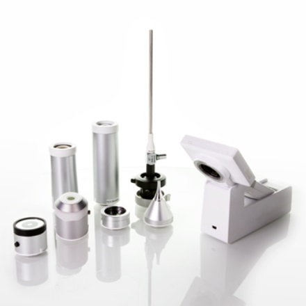
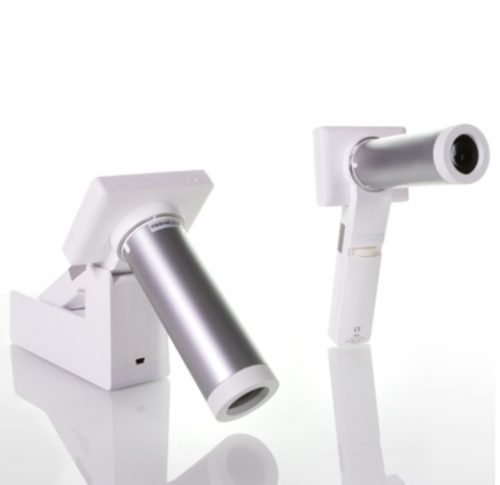
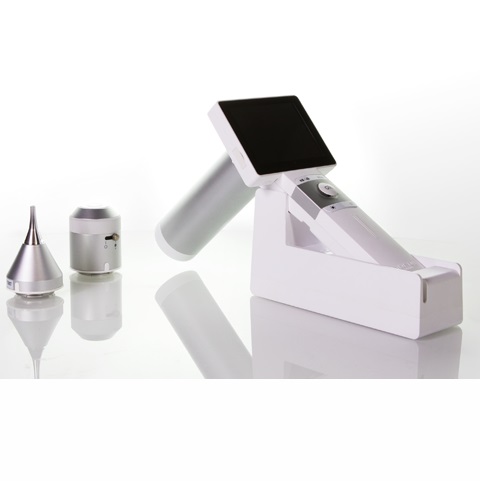 This innovative product “Digital Hand-held diagnostic set” is Class-II (ophthalmoscope)/Class-I (otoscope and dermoscope) Medical Device. It is expected to substitute for the traditional ophthalmoscope, otoscope and dermoscope by the use of the digital photographic solution. This medical device is provided to capture the digital photograph or video of eye-fundus, ear canal and tympanic membrane, epidermis and dermis of skin. Following the global trend of electronic medical records and telehealthcare networks, this people-oriented medical imaging product will be widely applied in doctors’ offices, clinics, skilled nursing facilities and healthcare station.
 -
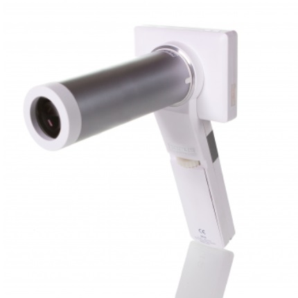
- Avoid unnecessary mydriatic medications and side effects.
- Improve quality and efficiency of making the rounds and even reach those in remote areas.
- Minimize paperwork with images or videos taken, stored, and processed digitally.
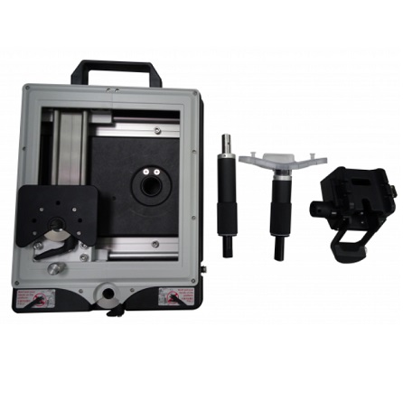
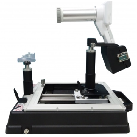
- Acquire additional stability with a special adapter attached to slit lamp.
-
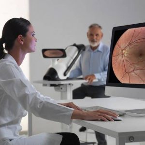
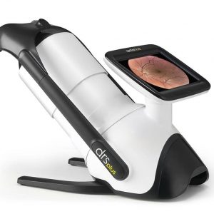
iCare DRSplus TrueColor confocal fundus imaging system
Key features- TrueColor Confocal Technology
- Multiple imaging modalities including red-free, external eye and stereo view imaging
- 2.5 mm minimum pupil size
- Fast, easy and fully automated operations
- Mosaic function which creates retinal panoramic views up to 80°
- Remote Viewer that allows for reviewing from devices on the same local area network
- Remote Exam feature enables executing an exam from a distance
-
-
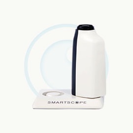
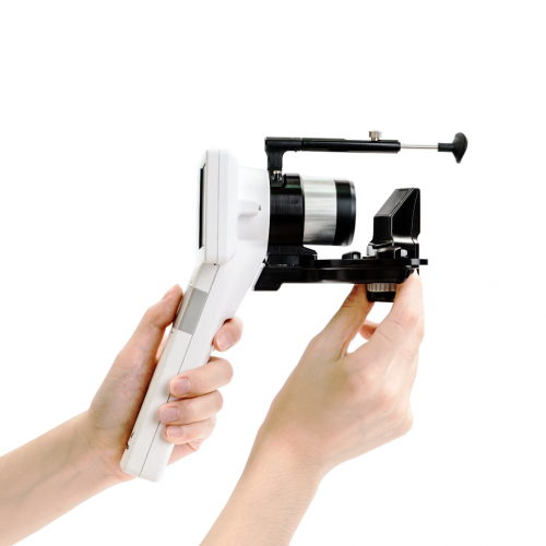
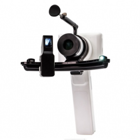
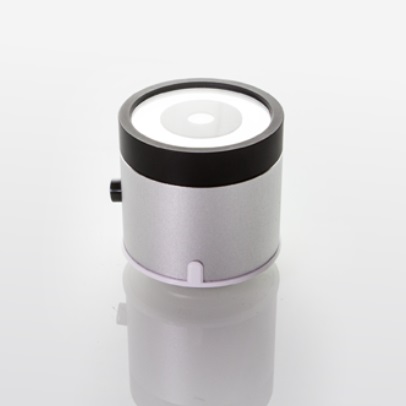
 Smartscope PRO camera with Smartscope FA module is a perfect combination of Slit Lamp and hand-held mode providing high quality Fluorescein Angiograms with a wide 40 degrees field of view. 9 internal fixation targets enable peripheral imaging (total FOV 70 * 52 degrees). Smartscope FA is easy to operate and offers fast image capture ability and a detailed view of the entire fluorescein dye circulation dynamics.
With a special Slit Lamp Adapter Smartscope FA can be mounted to any Slit Lamp with patient head rest.
Wi-Fi enables easy image transfer to PC, laptop, tablet or mobile devices. With Optomed Workstation software (sold separately) the images can be viewed, shared and stored locally and sent directly to a DICOM compatible system (PACS or other) of the hospital network.
 -
-
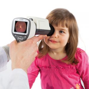
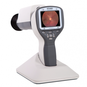
 Optomed Smartscope® PRO is the next generation hand-held medical camera that provides high image quality which fulfills international ISO 10940 fundus camera standard requirements. Interchangeable modular optics make the camera even more versatile. The lightweight camera incorporates advanced technology in a portable design.
Smartscope PRO camera provides accurate and silent autofocus which makes image capturing quick and easy. The camera is also equipped with Wi-Fi for advanced image transfer to any PC or mobile device.
The Smartscope PRO comes with Patient Data Management software (sold separately) with full DICOM and PACS compliance to ensure seamless interoperability with hospital networks and practice management systems.
 -
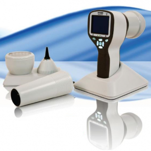
 Portable Non-mydriatic eye fundus examination
Optomed Smartscope M5 is a digital medical camera that provides general, ophthalmoscopic, otoscopic and dermatoscopic imaging with one hand-held device. This multipurpose digital imaging device weights only 400g and powered with a battery it gives you the freedom to move around and take the device with you to any location.
Digital still and video images created with Optomed Smartscope M5 allow making accurate first diagnosis and planning consistent follow-up treatment. Optomed Smartscope M5 is easily adopted into daily examination routines and connectivity to any patient database system enables fluent image data sharing e.g. for consultation purposes. The device comes with a 2GB SD memory card and pictures can be easily transferred to your PC by placing the device to its cradle which connects to the PC via USB connection.
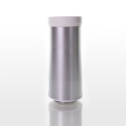
The 5 megapixel sensor and up to 2560 x 1920 pixels resolution guarantee the quality of your pictures. Each of our 4 attachable optics modules has its own light source and adjustable illumination levels. Optics are fast and easy to attach and detach with bayonet connectors.
The advances in M5 compared to M3-2 are new sensor enabling improved picture quality, higher battery capacity and keyboard lights that make using the device in a dark room easier.
Optomed Smartscope M5 offers non-mydriatic fundus imaging without the need for dilation drops with EY2 and EY3 optics modules.
 -
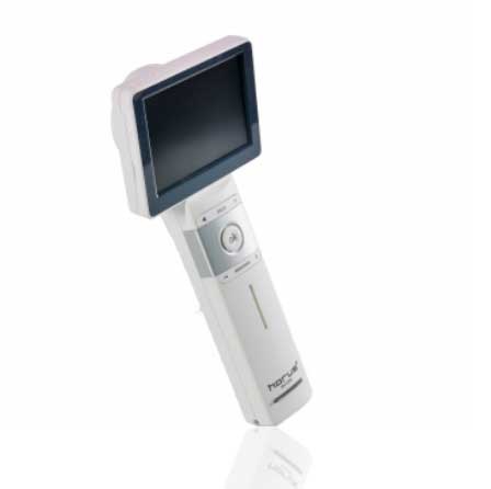
- Provide high definition clinical image
- Portable and hand held application in disease screen
- Friendly user interface with touch screen and Auto focus
- Multi functional diagnosis in ophthalmology , ENT . Dermatology and general practice.
- Widely application in clinic room , private office ,hospital, Tele- medicine , and mobile health.
-
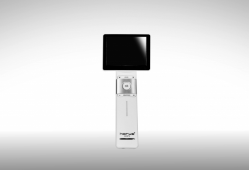
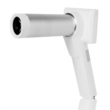 Horus DEC200 Non-Mydriatic Digital Handheld Fundus Camera offers high image quality with ISO 10940 fulfillment. 5MP (2592*1944 pixels) and 45 degree FOV of fundus image are captured to provide more details. 7 internal fixation targets for macula center, disk center and peripheral image in DEC 200 optical modules. Horus DEC200 provides both auto-focus and power-focus function to facilitate image capturing. Touch LCD Screen and Wi-Fi compatibility are also equipped . With a special slit lamp jig, Hours DEC 200 can be mounted with slit lamp for desktop application.
 -
