-
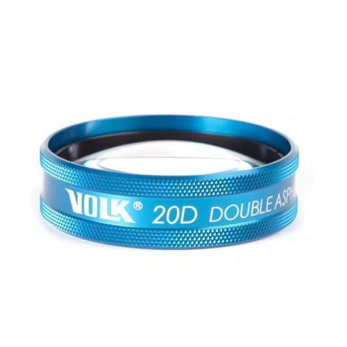
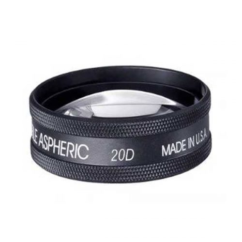 PART #V20LC Perhaps the most recognized lens around the world, the Volk 20D is the lens that started the legacy of double aspheric lens design for BIO and is still considered as a gold standard. The contributing factors for this are the perfect balance of field of view and magnification of the 20D. The working distance of 50 mm also makes lens manipulation a comfortable experience for the examiner. This lens is perfect for general diagnosis of patients with a 3x magnification and field of view that allows visualization up to the mid peripheral region. The dynamic examination with the appropriate eye movements by the patients allows viewing of the peripheral retina as well as detailed examination by BIO in the primary position provides a comprehensive estimate of retinal health. Thus, this lens is a great choice both as a first line of diagnosis as well as a high level central retinal examination.
 -
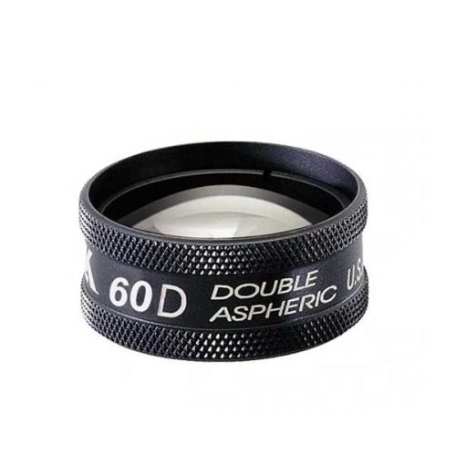 PART #V60C The 60D lens is one of the high magnification fundoscopy lenses used for a thorough and detailed examination of the central retina such as the macula and the nerve head. The trademark double aspheric design provides excellent detail and imaging for detecting subtle details and indications of retinal abnormalities. The optical profile of this lens requires a longer working distance of 18 mm from the patient. The high magnification provided by this lens is a great choice for diagnosis and assessment of the severity of Age-Related Macular Degeneration, following cup to disk ratios in patients and capillary hemorrhages. Dilation is required to obtain optimum retinal imaging with the 60D.
 -
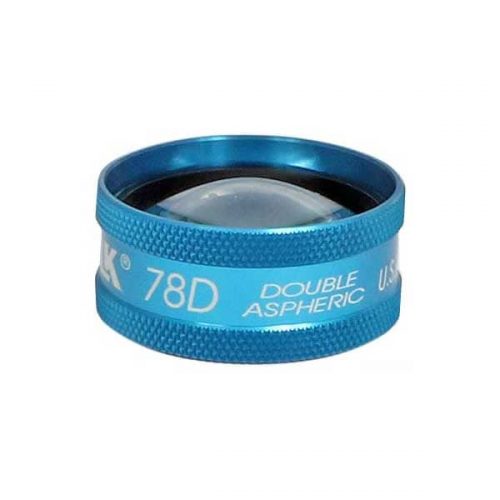
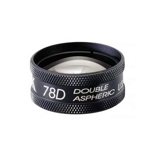 PART #V78LC The is an ideal lens for doctors who regularly cater to populations prone to glaucoma and other posterior pole abnormalities. The double aspheric design and field of view offered by a 78D offers clear and large views of the central mid-retinal regions. This lens offers a high magnification without cutting down too drastically on the field of view. This lens is a popular choice as a general diagnosis lens among doctors who prefer a larger ring size than the more common and smaller profile of the 90D. Dilation is required to obtain optimum retinal imaging with the 78D.
 -
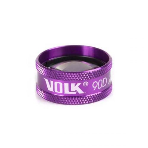
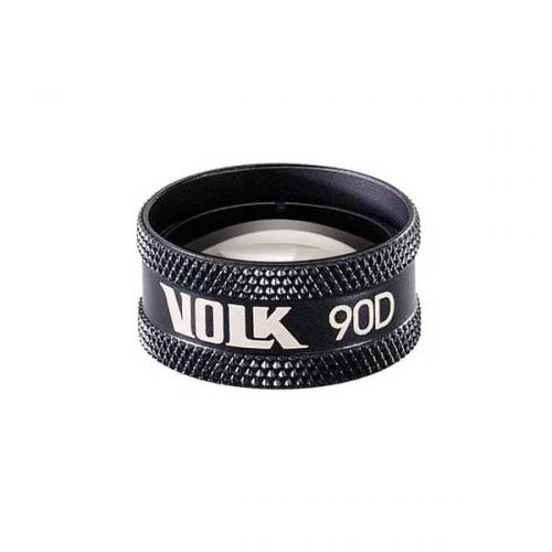 PART #V90C The Volk 90D is the most widely known fundoscopy lens and is to date considered the gold standard in exam rooms across the world. The small profile of the 90D coupled with its optical profile makes it a great first lens. This lens is a perfect choice for general examination and retinal imaging. This lens can be used to get through small pupils for patients who do not prefer or accommodate dilation and as a result is also a popular choice for undilated retinal exams.
 -
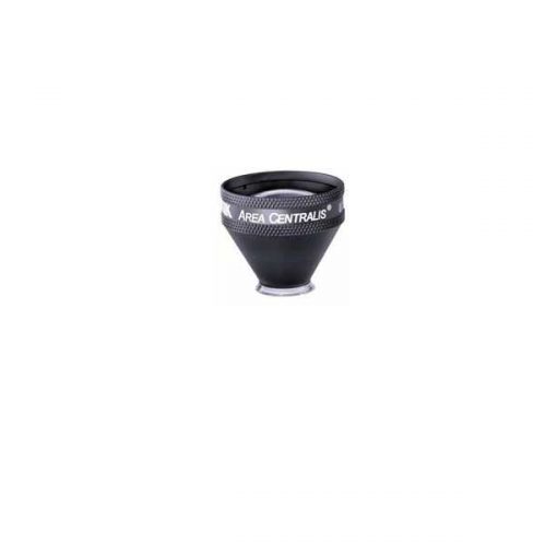 PART #VAC The Area centralis is designed with a 1.06x magnification to provide high detail, magnified views of the posterior pole. This lens is ideal for grid/focal laser procedures of the central retina for treating microaneurysms and edema in conditions such as diabetic retinopathy.
- High magnification for detailed examination of the posterior pole
- Available in flange, no flange and ANF+ contact options
- Ideal for focal/grid laser therapy
-
 PART #VDGTLCM The digital clear mag is designed specifically for high magnification and detailed examination of the macula and optic disc with a 3.89x magnification. This lens is an easy transition for those who are used to handling a 20D lens with a similar lens ring size and working distance but with an added advantage of high magnification. This lens is a great choice for detecting and monitoring the subtle changes in the optic disc morphology in patients with diabetic retinopathy, glaucoma and age related macular degeneration.
 -
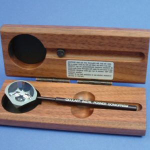
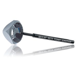 Ideal for use with children or patients with a small palpebral fissure. The instrument consists of a highly polished truncated silver surfaced pyramid with a plane anteroir viewing surface over four mirrors. An aluminum handle set at 35 degrees is bonded to one corner of the lens. The lens is used in the diamond position (45 degrees) resulting in fewer adjustments of lowering and elevating the slit beam with either hand. Item #: OCIOPDSG
 -
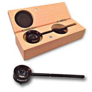 This 4 mirror glass lens is used for viewing all quadrants of the anterior chamber without lens repositioning. Mirrors are placed at angles of 62 degrees. No flange. No fluid required. Removable handle screws into place. Lens diameter: 20mm Rim Diameter: 25mm Lens Contact Point: 9mm Comes with padded case Item #: AMPDSG
 -
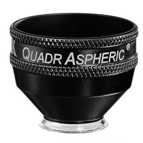 With a 144° field of view, this lens enables wide field visualization up to the peripheral retina for diagnosis and treatment of peripheral retinal defects. This lens is specially designed to provide wide field visualization even through small pupils. This application is critical when evaluating and treating patients such as those at risk of angle closure, neovascularization of the iris etc. in whom dilation should be avoided. The small pupil capability is also advantageous when treating geriatric populations in whom pupil response to dilation is limited. The large flange on this lens provides the perfect stability and control over the eye needed during laser procedures
- Wide-field distortion free viewing of the retina
- Small-pupil capability
- Ideal for detecting and treating mid to peripheral retinal abnormalities
- Available in Flange, no Flange and ANF+ contact options
-
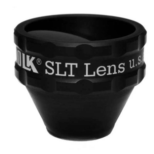 PART #VSLT This lens is a classic one mirror design lens that provides a large, clear view of the angle structures for performing SLT procedures to lower intraocular pressure in patients with ocular hypertension and glaucoma. This lens is designed for compatibility when used with a frequency doubled Q switched Nd:YAG laser. The lens has to be rotated gently on the patient’s eye to aim the laser at the various sections of iridocorneal angle. The 1.0x magnification of this lens helps maintain the laser spot size and density during laser delivery.
- Selective Laser Trabeculoplasty (SLT)
- Ideal for Glaucoma Treatment
- Laser Trabecular Meshwork treatment
-

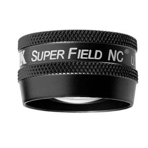 PART #VSFNC Often referred to as a ‘Super 90D’ this lens provides wide field imaging out to the mid periphery and dynamic viewing out to the periphery. This lens is a great choice for pan-retinal examination. This lens provides the magnification of a 90D, with an increased field of view enabling high resolution imaging of the posterior pole. This combination of magnification and wide field imaging allows quick and comprehensive examination of the retina, making it a go-to lens for general examination. A 30 mm lens ring provides a comfortable grip and manipulation of the lens within the orbit.
 -
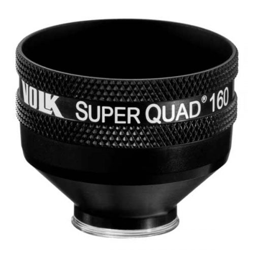 PART #VSQUAD160 Experience wide field, distortion-free visualization of the retina from the nerve head and macula, up to the ora serrata, designed for detection and treatment of retinal abnormalities like peripheral retinal tears, peripheral retinal detachments, giant retinal tears etc. The 30 mm lens surface offers a large, clear image of the retina for accurate and easy placement of the laser spot. The contact surface is designed carefully to provide optimum stability on the patient’s cornea while ensuring patient comfort.
- Wide-field distortion free viewing
- Ideal for detecting and treating mid to far-peripheral retinal abnormalities
- Large lens surface area providing a large working area
- Available in: Flanged contact, no Flange contact design


