- High magnification for detailed examination of the posterior pole
- Available in flange, no flange and ANF+ contact options
- Ideal for focal/grid laser therapy
-
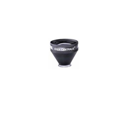 PART #VAC The Area centralis is designed with a 1.06x magnification to provide high detail, magnified views of the posterior pole. This lens is ideal for grid/focal laser procedures of the central retina for treating microaneurysms and edema in conditions such as diabetic retinopathy.
 -
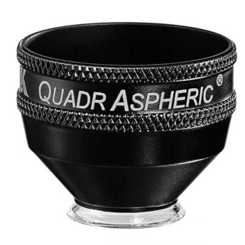 With a 144° field of view, this lens enables wide field visualization up to the peripheral retina for diagnosis and treatment of peripheral retinal defects. This lens is specially designed to provide wide field visualization even through small pupils. This application is critical when evaluating and treating patients such as those at risk of angle closure, neovascularization of the iris etc. in whom dilation should be avoided. The small pupil capability is also advantageous when treating geriatric populations in whom pupil response to dilation is limited. The large flange on this lens provides the perfect stability and control over the eye needed during laser procedures
- Wide-field distortion free viewing of the retina
- Small-pupil capability
- Ideal for detecting and treating mid to peripheral retinal abnormalities
- Available in Flange, no Flange and ANF+ contact options
-
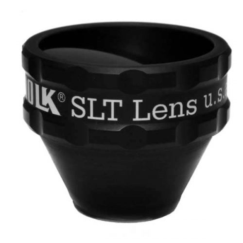 PART #VSLT This lens is a classic one mirror design lens that provides a large, clear view of the angle structures for performing SLT procedures to lower intraocular pressure in patients with ocular hypertension and glaucoma. This lens is designed for compatibility when used with a frequency doubled Q switched Nd:YAG laser. The lens has to be rotated gently on the patient’s eye to aim the laser at the various sections of iridocorneal angle. The 1.0x magnification of this lens helps maintain the laser spot size and density during laser delivery.
- Selective Laser Trabeculoplasty (SLT)
- Ideal for Glaucoma Treatment
- Laser Trabecular Meshwork treatment
-
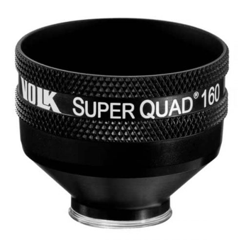 PART #VSQUAD160 Experience wide field, distortion-free visualization of the retina from the nerve head and macula, up to the ora serrata, designed for detection and treatment of retinal abnormalities like peripheral retinal tears, peripheral retinal detachments, giant retinal tears etc. The 30 mm lens surface offers a large, clear image of the retina for accurate and easy placement of the laser spot. The contact surface is designed carefully to provide optimum stability on the patient’s cornea while ensuring patient comfort.
- Wide-field distortion free viewing
- Ideal for detecting and treating mid to far-peripheral retinal abnormalities
- Large lens surface area providing a large working area
- Available in: Flanged contact, no Flange contact design

