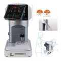
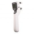
-
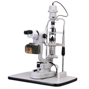 SLR camera (24.1 Mega Pixels); Special case storage system Professional image analyzing, processing and management system; Flash illumination compensation system High definition video function Background illumination system Power box supplies camera power directly. without charging battery Super optical Top definition eyepiece Adjusting the illumination by Joystick, it is much easier to use. Economic, high quality, low maintenance and depreciation New function: Illumination control by Joystick Eyepiece correction (Same focus) Automatic image optimization (such as color, brightness) Filter for corneal staining (Five different color is more easier for doctor to get best images)
 -
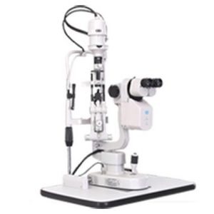 Professional image analyzing, processing and management system Professional optics with CCD Special case storage system Professional software with video function Background illumination system Flash function
 -
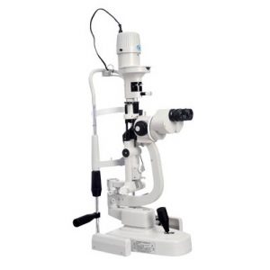
- Galileo parallel design for using, five magnifications 14mm aperture provides broader and clearer field of view High sensitivity professional objective lens German industrial bulb, high brightness without harsh, quick heat dispersion, also provide the low heating, high brightness and even lighting. International standard accessory interface which can connect kinds of ocular posterior segment laser adapter and foldman tonometer Digital upgrading interface. Can upgrade to digital slit lamp Excellent optics performance, versatility and ease of use Stereo variator, besretinal view even through small pupils Ergonomic, wide aperture optics for fatigue free daily use
-
 Multiple aperture/filter options, combined with halogen light provide long-lasting, reliable performance for general and specialist examinations. A versatile ophthalmoscope at an economical price.
 -
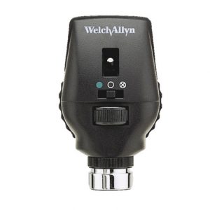 The patented Welch Allyn Coaxial Vision System facilitates ophthalmoscopy by enabling easier entry into the eye, a larger field of view, and reduced glare compared to standard ophthalmoscopes.
 -
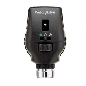 Patented Welch Allyn Coaxial Vision optics, combined with 68 lenses in single-diopter steps, for the precision you need to conduct a quality eye exam.
 -
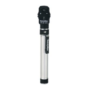 Patented Welch Allyn Coaxial Vision optics, combined with 68 lenses in single-diopter steps, for the precision you need to conduct a quality eye exam.
 -
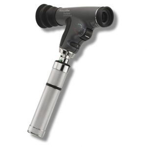
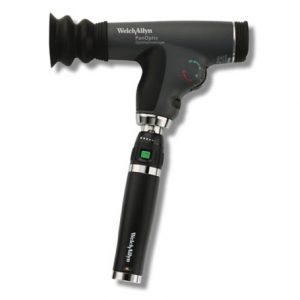 Our revolutionary PanOptic Ophthalmoscope addresses the fundamental challenge in ophthalmoscopy—to get a good view of the fundus in order to make a sufficient assessment. Patented Axial PointSource™ Optics make it easy to enter undilated pupils, offering a 25º field of view, resulting in a view of the fundus that's 5X greater than you see with a standard ophthalmoscope in an undilated eye. Direct viewing of the fundus through the PanOptic provides better images of the retinal changes caused by hypertension, diabetic retinopathy, glaucoma, and papilledema to enable clinicians to make these diagnoses earlier.
 -
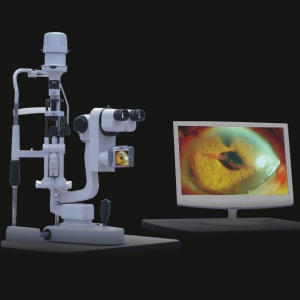 Features Binocular linkage Galileo style Five Steps Rotation magnification Large depth of field and high definition optical system Digital camera: image captured by button on handle Image system software based on Windows 2000/XP (optional)
 -
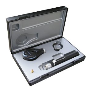
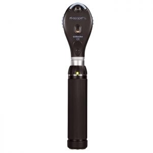 All ri-scope® L ophthalmoscopes with modular design impress with their high-performance optics with aspherical condenser lens with reduced-refl ection view even with small pupils and through improved ergonomics. The new ri-scope® L ophthalmoscopes have a thumbrest for securing the ophthalmoscope, which enables easy control of all the elements with one finger. ri-scope® L2 The enhanced basic model with separately engageable red-free, blue and polarization filters for each aperture.
- Diopter wheel with 29 corrective lenses
- Plus 1-10, 12, 15, 20, 40; Minus 1-10, 15, 20, 25, 30, 35
- Easy-to-operate aperture hand-wheel with semi-circle, small/medium/large circle, fixation star, and slit
- Includes filter wheel engageable for all apertures with symbol display, red-free filter, blue filter and polarization filter
- Optimized high-performance optics with aspherical condenser lens
- Parallel beam path
- Dust-proof
-
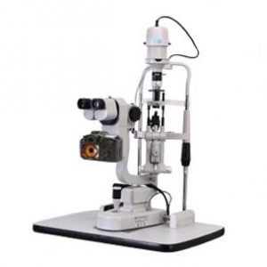 Professional image analyzing, processing and management system Professional optics with high definition digital SLR camera (16.2 Mega Pixels) Special case storage system Professional software with high definition video function Background illumination system

