-
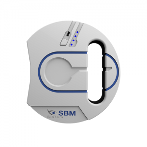
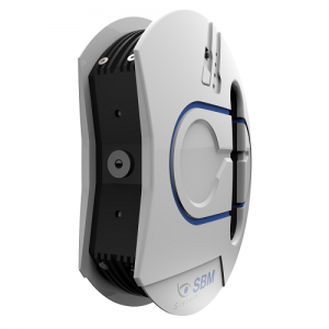 Typology: Device for evaluation of the meibomian glands Camera: Colored, sensitive to Infrared (NIR) Part examined: Upper and lower eyelids Graphic result: Coloration absent area and present Tools: Editor to highlight the area if the glands to be evaluated Resolution: 8 mp Light Source: Infrared LED Rating: Calculation of percentage of missing glands
 -
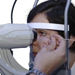
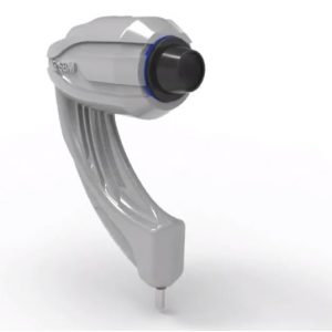 Acquisition Mode: Multi shot, tube, movie ISO Management: Variable Grids: Placido disc, NIBUT grid Light source: Infrared LED - Blue and White LED Image Resolution: 8 mp Focus: Autofocus, Manual focus Camera: Colored, Sensitive to Infrared (NIR)
 -
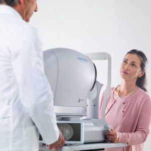
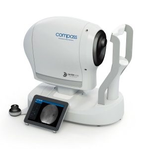
iCare COMPASS Automated Perimeter with active Retinal Tracking
Key features
- Standard automated perimetry
- Active retinal tracking compensating for poor patient fixation in real-time
- Auto-focus — no trial lens needed
- Hygienic design
- Illustrative fixation analysis; fixation area and plot
- High-resolution confocal TrueColor imaging of the retina
- No dilation of pupil needed, the patient can blink freely and the test can be suspended at any time without data loss
- Ease of use & minimal operator training
-
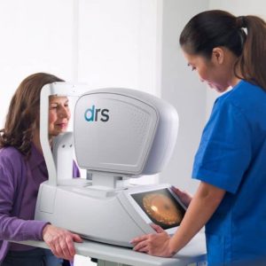
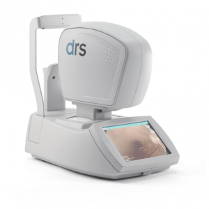
iCare DRS automated digital retinography system
Key features- Fully automated: patient autosensing, auto-alignment, auto-focus, auto flash adjustment, auto-capture
- Short exam time
- High quality color and Red-free images
- Minimal training required
- Compact and clean design
- No additional PC required
- Touch-screen operation
-
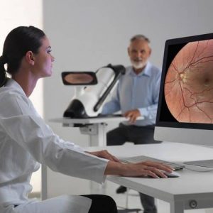
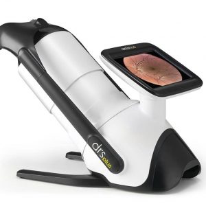
iCare DRSplus TrueColor confocal fundus imaging system
Key features- TrueColor Confocal Technology
- Multiple imaging modalities including red-free, external eye and stereo view imaging
- 2.5 mm minimum pupil size
- Fast, easy and fully automated operations
- Mosaic function which creates retinal panoramic views up to 80°
- Remote Viewer that allows for reviewing from devices on the same local area network
- Remote Exam feature enables executing an exam from a distance
-
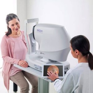
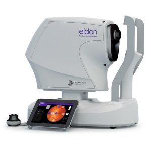
iCare EIDON widefield TrueColor Confocal fundus imaging system
Key features- Multiple imaging modalities including TrueColor, blue, red and Red-Free and infrared confocal images
- Widefield, ultra-high-resolution imaging
- Capability to image through cataract and media opacities
- Dilation-free operation (minimum pupil 2.5 mm)
- Flexibility of fully automated and fully manual mode
- All-in-one compact design, no additional PC required
-
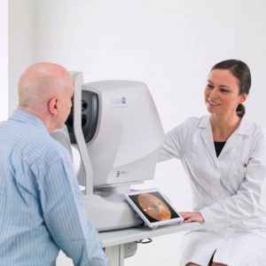
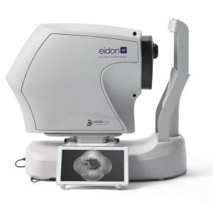
iCare EIDON AF blue autofluorescence confocal fundus imaging system
Key features
- TrueColor, blue autofluorescence, infrared and RGB channels confocal images
- Panoramic view of the retinal autofluorescence (up to 110°)
- High details and contrast in the same device
- Short exam time and enhanced patient comfort
- Easy to use, speeds up patient workflow
-
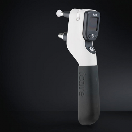
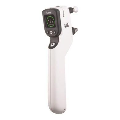
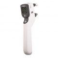
iCare IC200 tonometer - introducing a new era in clinical tonometry
Key features
- 200-degree position freedom
- Suitable for all patients
- Consistent and accurate readings
- No anesthetic drops
- Improved probe control
- User interface in multiple languages
- Wireless connection to iCare EXPORT
- Wireless printing
- No calibration
-
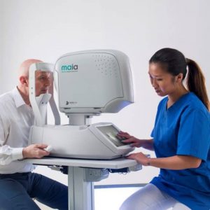
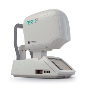
iCare MAIA and S-MAIA confocal microperimeters
Key features- 25 Hz eye-tracking with confocal SLO imaging
- Pre-programmed and custom tests
- Sensitivity and fixation indexes, illustrative sensitivity, and fixation maps
- Informative examination and progression reports
- 2.5 mm minimum pupil size
- Patients with cataract (up to grade 3+) and media opacities can be examined
- Auto-focus (from -15D to +10D)
- Sensitive to functional changes due to macular pathologies or treatments even in early stages (background down to <0.0001 asb, threshold range 36 dB)
- Simple to use, patients can be tested in less than 3 minutes per eye
- S-MAIA includes also scotopic testing with cyan and red stimuli
-
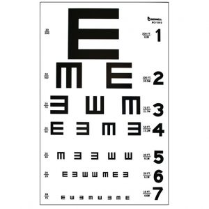 9" x 14" TRANSLUCENT PLASTIC MAY BE USED WITH ILLUMINATED TEST CABINET OR AS A WALL CHART 20/200 to 20/15. Item #: BC1262
 -
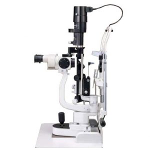 The L-0189 5 STEPS MAGNIFICATION SLIT LAMP provides practitioners with the utmost satisfaction thanks to its excellent image clearness and fine design.
 -
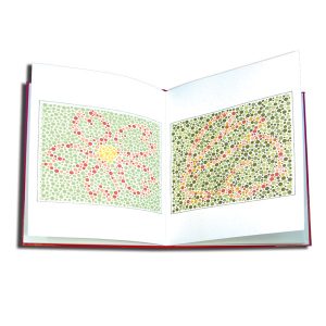 This test is similar to the Ishihara Isochromatic Test. 10 plates with non-verbal animal patterns. Originally created by Hiro Matsubara, MD. Printed in Japan. Item #: AT400


