-
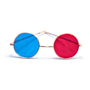 Reversible Metal Frame Glasses with Red/Blue Lenses These glasses are convenient for testing or training. Use with Tranaglyphs to alternate demand. Item #: BC1100RB
 -
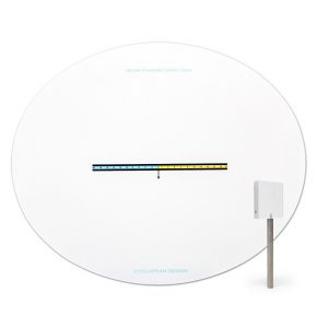
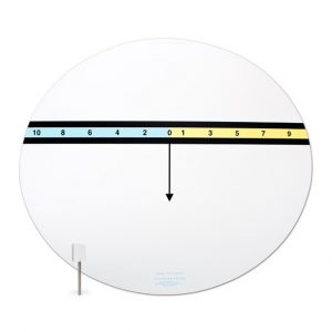 Both Near and Far come with a single 6 PD stick prism. These cards allow direct measurement of the distance and near phorias in real space or with a phorpter. The patient simple has to tell which number the arrow points to. Any odd number (in yellow) shows esophoria while the even numbers (in blue) shows exophoria. An AC/A can quickly be determined with trial lenses. The reverse side of the near card has a X-Cyl target, and a variety of letter and word targets. Near card is about 12" wide. Distance chart is about 24" wide. Item #: CDHP+
 -
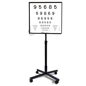 The ESC2000 Illuminator Cabinet provides a uniform retro-illuminated testing surface, ideal for clinical testing of high contrast and low contrast LogMAR tests, including ETDRS, Lea Symbols, Lea Numbers, Landolt C, etc. The cabinet utilizes a pure-white LED light source. (Stand not included. Can be purchased separately. Part # GL50004)
 -
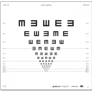 Illiterate Tumbling E ETDRS vision chart with proportionally spaced (geometric progression) lines; line sizes range from 20/200 to 20/10 (6/60 to 6/3) equivalent.
 -
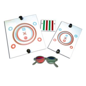
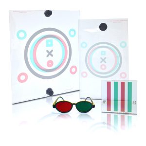 Red/Green TV trainer or can be placed on a printed page and used as a reading unit. Used to build binocularity in true space.
 -
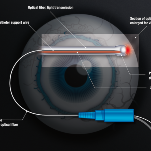
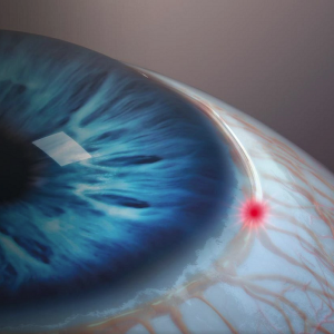
The optimum in minimally invasive, canal-based glaucoma surgery
With a mild touch and manifest efficacy, ABiC™ performed with the iTrack™ surgical system is a comprehensive minimally invasive glaucoma surgery that can effectively reduce IOP and eliminate or reduce the medication burden. Restorative and atraumatic, ABiC™ can be performed across the entire glaucoma disease process – and in conjunction with other treatments and MIGS options. -
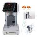
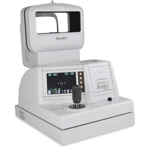 Natural Vision Auto Ref-Keratometer Natural Vision Auto Ref-Keratometer NVISION-K5001 is a unique Auto Ref-Keratometer that allows the patient to look through a very wide window. This feature helps the patient relax during the measurement as looking through a window with both eyes can minimise instrument accommodation.
 -
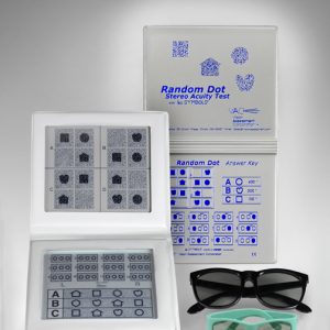 Designed to rapidly test for amblyopia and strabismus in early and non-readers and non-verbal children & adults. Expanded Random Dot LEA Symbols Test (500, 250, 125, 63 seconds of arc). Graded circle test now down to 12.5 seconds with no monocular clues. New improved booklet has answer key on back cover and includes polarized viewers.
- Expanded Random Dot Lea Symbols Test
- Graded circle test now down to 12.5 seconds
- No monocular clues with NEW technology
- New improved booklet
- Answer key on back cover
- Includes both adult and pediatric polarized viewers
-
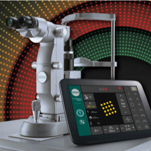
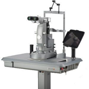
 Integre PRO Scan combines multi-color photocoagulation with precise computer-controlled pattern scanning in our unique all-in-one laser/slit lamp design.
A Pattern and Wavelength for Every Pathology
Whether accurately positioning focal treatment in the macular area, or performing PRP in the periphery, Integre Pro Scan™ provides a pattern and wavelength for every pathology. The system offers a comprehensive array of pattern combinations: shape, size, and rotation can be fully customized in order to best meet your clinical requirements. Patterns of up to 16 spots can be delivered sequentially, as well as single spots. In addition, the system’s computer-controlled operation ensures precise pattern spacing and consistent shaping of laser spots and patterns for more accurate, consistent treatment results. Integre Pro Scan™ also provides a number of wavelength configurations, including two dual-wavelength configurations. This includes a high-power yellow and red configuration that delivers the full treatment spectrum of a traditional three-color multi-color photocoagulator.- yellow-red configuration (561 nm and 670 nm)
- green-red configuration (532 nm and 670 nm)
- green configuration (532 nm)
- yellow configuration (561 nm)
Predictable Treatment Outcomes
A proprietary ZenTec™ laser cavity delivers uniform energy distribution across the full diameter of the spot, eliminating hotspots and achieving optimal homogenous burns – delivering consistent, predictable treatment outcomes over a number of different operating conditions.Designed to Maximize Your Workflow
Unlike other photocoagulator systems, which require an external dichroic or fixed mirror in order to connect the laser to the slit lamp, Integre Pro Scan’s unique, all-ine-one laser/slit lamp design channels the laser directly through the slit lamp optics. The end result is better visualization and optimal illumination. In addition, this integrated design minimizes system downtime, because there are no exposed fiber optic or electrical cables. -
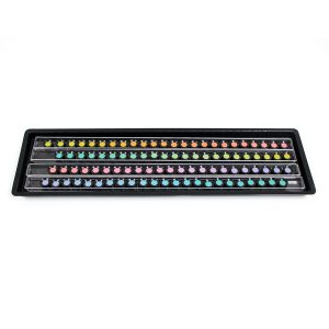 This handheld circular pupillometer device is used for measuring different size pupils. It was developed and researched by Dr. Jack Richman, O.D., as part of the Massachusetts Drug Evaluation and Classification Program (MDEP). This pupil measurement device is used by law enforcement officers across the country to assist in determining whether people are possibly impaired and/or under the influence of drugs. The reverse side has expected pupil size in different lighting conditions for non-impaired people. Item #: LV3553000
 -
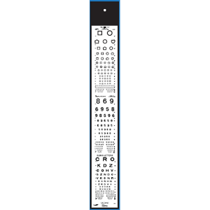 Designed for use in manual Chart Projectors including products from: R.H. Burton, Marco, Reichart, Topcon, and Woodlyn. Includes test for: → LEA Symbols® 20/100 – 20/10 (18 Lines) → LEA Numbers® (16 Lines) (Multiple Lines 20/50 – 20/10) → Sloan Numbers 20/100 – 20/10 (16 Lines) (Multiple Lines 20/50 – 20/10) Item #: VA3202
 -
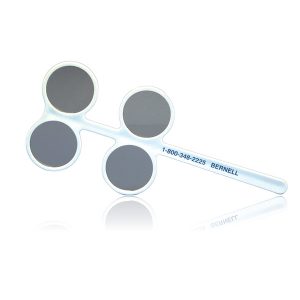 Four-well flipper bar features two sets of lenses to alternate demand. Used with Tranaglyphs or Vectograms to reverse BI/BO demands. Item #: BC1270POL


