-
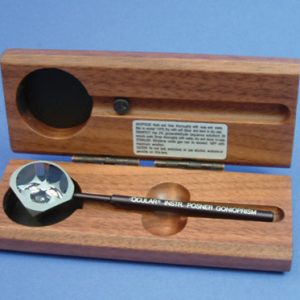
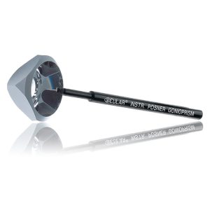 Ideal for use with children or patients with a small palpebral fissure. The instrument consists of a highly polished truncated silver surfaced pyramid with a plane anteroir viewing surface over four mirrors. An aluminum handle set at 35 degrees is bonded to one corner of the lens. The lens is used in the diamond position (45 degrees) resulting in fewer adjustments of lowering and elevating the slit beam with either hand. Item #: OCIOPDSG
 -
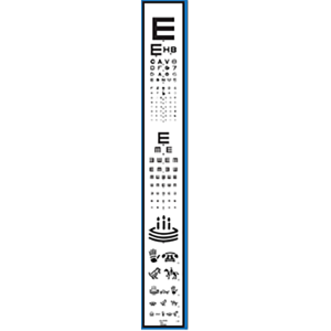 Designed for use in manual Chart Projectors including products from: R.H. Burton, Marco, Reichart, Topcon, and Woodlyn. Includes test for: → Sloan Letters 20/400 – 20/15 (15 Lines) → Tumbling E 20/200 – 20/25 (9 Lines) → Allen Acuity 20/200 – 20/30 (6 Lines) Item #: VA1192
 -
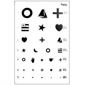 9" x 14" Translucent Plastic May be used with illuminated test cabinets or as a wall chart 20/100 to 20/10 Item #: BC1263
 -
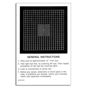 Refined Central Amsler Grid Amsler grid pad with additional lines at the central fixation point for testing and monitoring macular problems. Pad of 50 sheets
 -
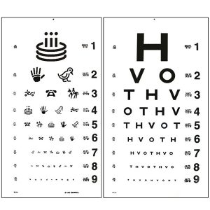 This test for illiterate patients has HOTV letters on one side and Allan figures on the other. Can be used with HOTV near card for matching with non-verbal patients. 21.5" x 11.5"
 -
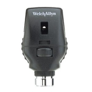 Multiple aperture/filter options, combined with halogen light provide long-lasting, reliable performance for general and specialist examinations. A versatile ophthalmoscope at an economical price.
 -
 Anaglyphic filters in primary colors
 -
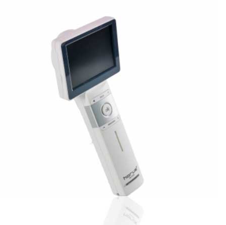
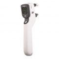
- Provide high definition clinical image
- Portable and hand held application in disease screen
- Friendly user interface with touch screen and Auto focus
- Multi functional diagnosis in ophthalmology , ENT . Dermatology and general practice.
- Widely application in clinic room , private office ,hospital, Tele- medicine , and mobile health.
-
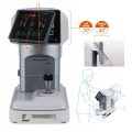
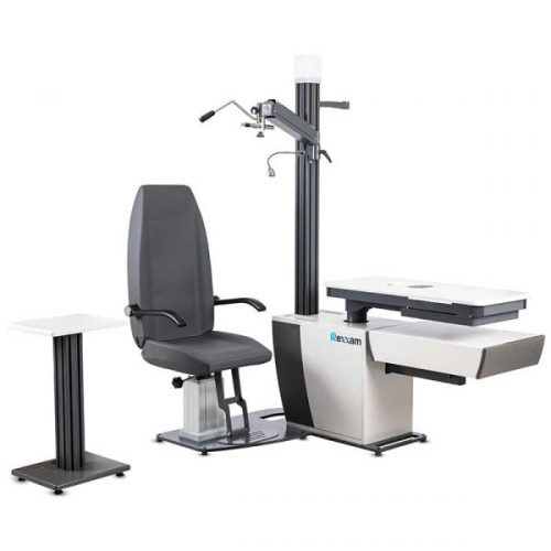 Ophthalmic unit Rexxam is introducing Our State-of-the-Art Ophthalmic Unit: Rexxam WorkStation OZY which can accommodate two instruments on a sliding rotatable tabletop. Embark on a new era of innovation, precision and comfort in eye care with our latest addition – the cutting-edge Ophthalmic Unit WorkStation OZY. Meticulously crafted to enhance the diagnostic and examination capabilities of our esteemed opticians and optometrists, this advanced ophthalmic unit is set to redefine the standards of patient comfort, operability and flexibility in eye-care.
 -
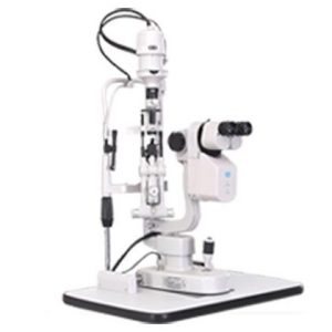 Professional image analyzing, processing and management system Professional optics with CCD Special case storage system Professional software with video function Background illumination system Flash function
 -
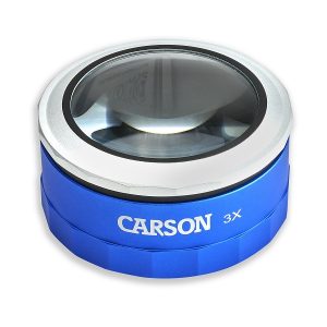 The MT-33 MagniTouch is a 3x power touch activated loupe. It has 3 super bright LED lights and a crystal clear glass lens. Use the on/off switch for “steady” light setting. The twist-action focus allows you to “zoom-in” for greater detail. The MagniTouch is a great Magnifier for detailed inspection, to view stamps and coins and for reading fine print or as a desk accessory. Uses 2 CR2016 batteries (included).
 -
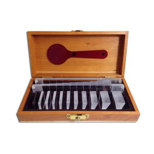 Basic prism set in handsome wood case, foam-lined for protection. Item #: APS30+
+65 6514 0848|info@ophthalmic.com.sg