-
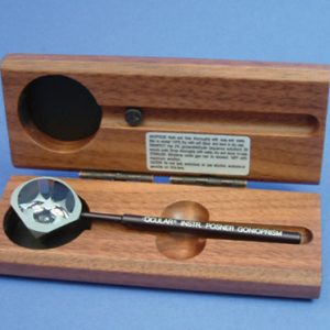
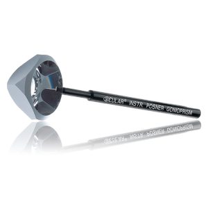 Ideal for use with children or patients with a small palpebral fissure. The instrument consists of a highly polished truncated silver surfaced pyramid with a plane anteroir viewing surface over four mirrors. An aluminum handle set at 35 degrees is bonded to one corner of the lens. The lens is used in the diamond position (45 degrees) resulting in fewer adjustments of lowering and elevating the slit beam with either hand. Item #: OCIOPDSG
 -
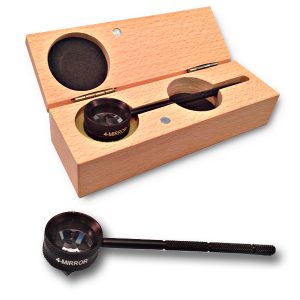 This 4 mirror glass lens is used for viewing all quadrants of the anterior chamber without lens repositioning. Mirrors are placed at angles of 62 degrees. No flange. No fluid required. Removable handle screws into place. Lens diameter: 20mm Rim Diameter: 25mm Lens Contact Point: 9mm Comes with padded case Item #: AMPDSG
 -
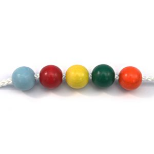 Some doctors prefer the size and feel of these string units over the Bernell ones above. These have Red, Green, Yellow, Blue, and Orange beads. Item #: FR+
 -
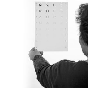
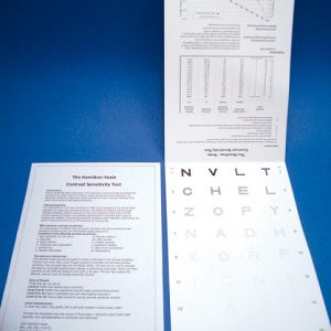 Hamilton-Veale Contrast Sensitivity Test This easy and convenient test can be used to monitor a decrease on the contrast sensitivity function over time. This test also demonstrates the difficulty of seeing in poor lighting or at night.
 -
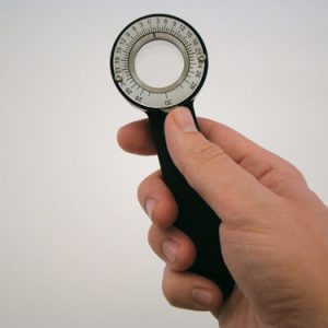
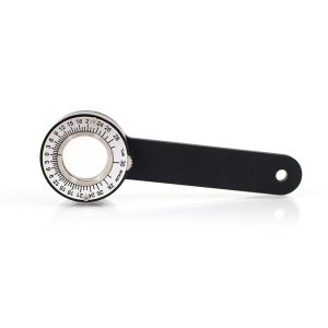 An affordable Risley Prism that you can use for testing. This prism allows variable prism demand up to 30PD. A prism before each eye can increase the demand of 60PD. The prism is approximately 1" diameter. Item #: ARPHH30
 -
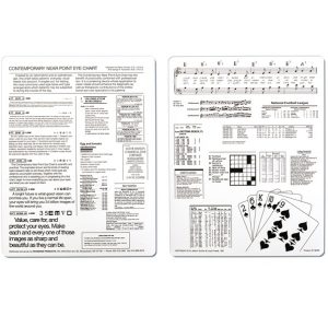 This sturdy 8.5" x 11" chart was created by an MD and OD to show most common task using your eyes. It can be used to screen for visual difficulties as well as demonstrate needs. Good for use in the exam room as well as the workup and dispensing rooms.
 -
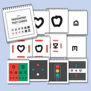 Collection of 66 test cards for use during examination of children. Hanks Pediatric Optotypes - Calibrated from 6/60 to 6/4. Designed for this purpose and tested in clinical trials to confirm recognition of the symbols by young children. Landolt C & Illiterate E Optotypes are also included - Calibrated from 6/60 to 6/4. Color Vision Screening - Pseudo-isochromatic plates with letters, numbers, circles and trails. Quality Production - Each card is matt laminated and encapsulated for long life protection. Actual size of each card is 108 x 112mm. Item #: AUPTC
 -
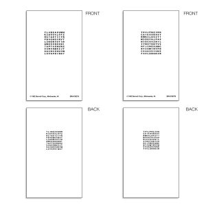
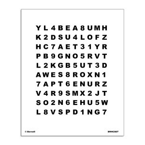 Hart Chart™ Accommodative Rock Chart Set Near and Far Chart Set provides letter and number matrix for testing and accommodative vision training. This is the classic Hart chart used by professionals for years.
 -
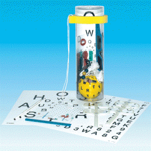 Home Trainer Tube Kit (VTE) 30 day lead time Tube designed to hold a selection of home training products. The plastic transparent tube can be personalized by printing logo and instructions in the paper cover.
 -
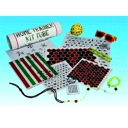 Home Trainer Kit Tube (VTE) for Children The tube contains a selection of products for children's home VT.
 -
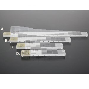
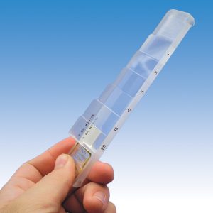 A.) "COMPLETE BAR" Includes prisms: 1, 2, 4, 6, 8, 10, 12, 14, 16, 18, 20, 25, 30, 35, 40 & 45 B.) "EXTENDED BAR" Includes prisms: 1, 2, 4, 6, 8, 10, 12, 15, 20 and 25. C.) "BETTER BAR" Includes prisms: 1, 3, 5, 10, 15 & 20. D.) "GOOD BAR" Includes prisms: 3, 5, 10, 15 & 20. Item #: AHB+
 -
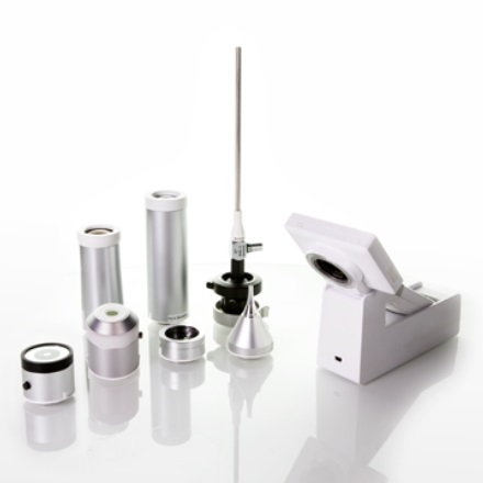
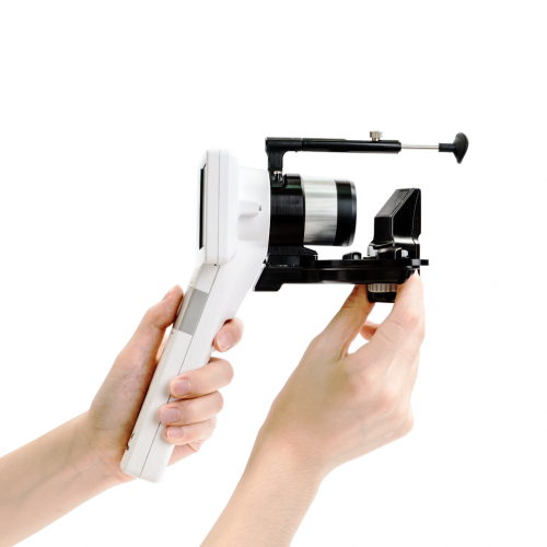
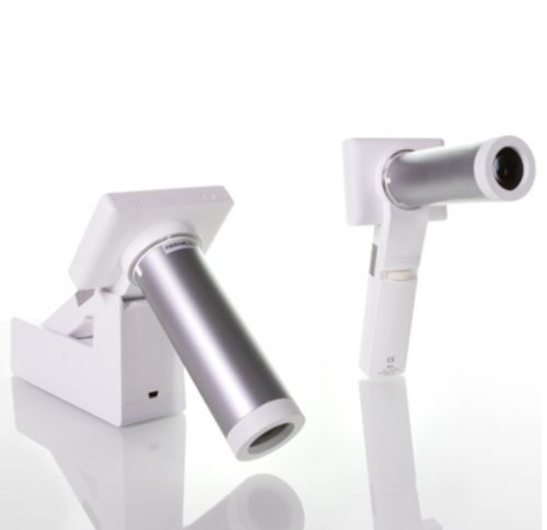
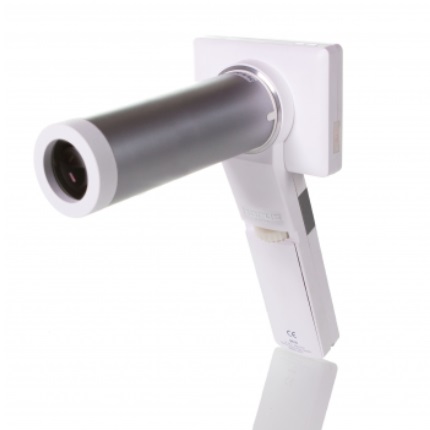
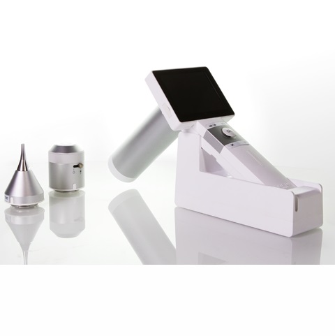 This innovative product “Digital Hand-held diagnostic set” is Class-II (ophthalmoscope)/Class-I (otoscope and dermoscope) Medical Device. It is expected to substitute for the traditional ophthalmoscope, otoscope and dermoscope by the use of the digital photographic solution. This medical device is provided to capture the digital photograph or video of eye-fundus, ear canal and tympanic membrane, epidermis and dermis of skin. Following the global trend of electronic medical records and telehealthcare networks, this people-oriented medical imaging product will be widely applied in doctors’ offices, clinics, skilled nursing facilities and healthcare station.
 -
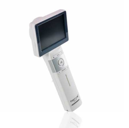
- Provide high definition clinical image
- Portable and hand held application in disease screen
- Friendly user interface with touch screen and Auto focus
- Multi functional diagnosis in ophthalmology , ENT . Dermatology and general practice.
- Widely application in clinic room , private office ,hospital, Tele- medicine , and mobile health.
-
-
- Avoid unnecessary mydriatic medications and side effects.
- Improve quality and efficiency of making the rounds and even reach those in remote areas.
- Minimize paperwork with images or videos taken, stored, and processed digitally.
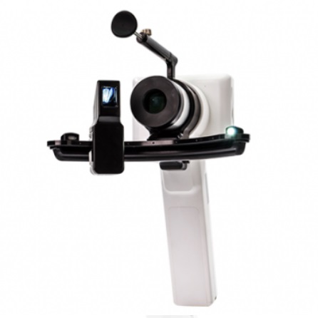
- Acquire additional stability with a special adapter attached to slit lamp.
-
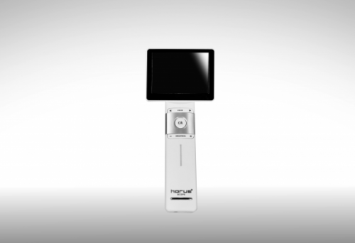
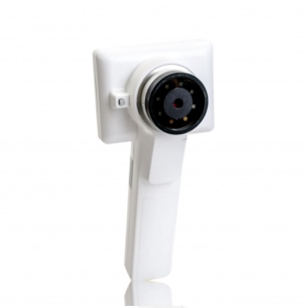
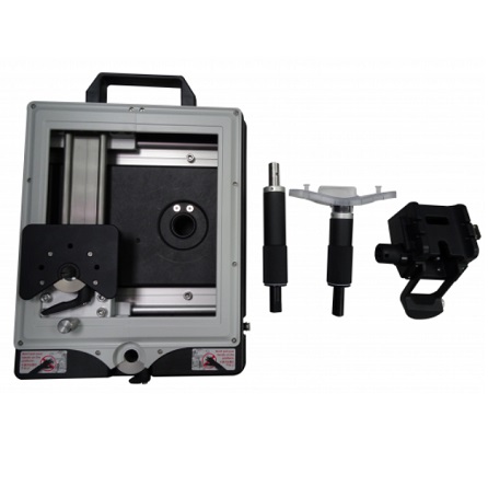
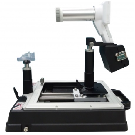
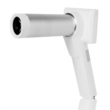 Horus DEC200 Non-Mydriatic Digital Handheld Fundus Camera offers high image quality with ISO 10940 fulfillment. 5MP (2592*1944 pixels) and 45 degree FOV of fundus image are captured to provide more details. 7 internal fixation targets for macula center, disk center and peripheral image in DEC 200 optical modules. Horus DEC200 provides both auto-focus and power-focus function to facilitate image capturing. Touch LCD Screen and Wi-Fi compatibility are also equipped . With a special slit lamp jig, Hours DEC 200 can be mounted with slit lamp for desktop application.
 -
-
-
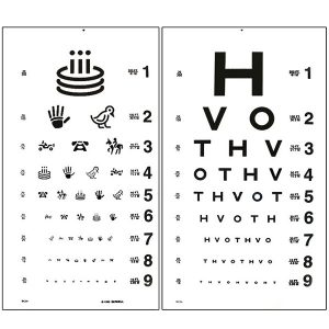 This test for illiterate patients has HOTV letters on one side and Allan figures on the other. Can be used with HOTV near card for matching with non-verbal patients. 21.5" x 11.5"
 -
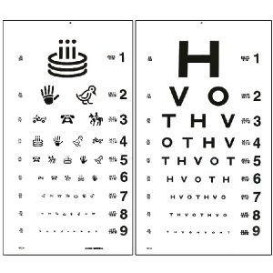 This test for illiterate patients has HOTV letters on one side and Allan figures on the other. Can be used with HOTV near card for matching with non-verbal patients. 21.5" x 11.5"
 -
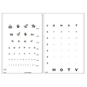 This card is ideal for testing illiterate patients. It has HOTV letters on one side and Allen figures on the other. CARD SHOULD BE HELD AT 16" 5" x 8" BCNIE comes with 16" elastic cord to verify testing distance.
 -
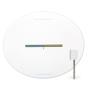
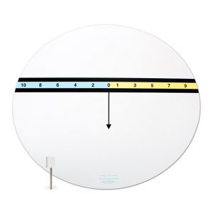 Both Near and Far come with a single 6 PD stick prism. These cards allow direct measurement of the distance and near phorias in real space or with a phorpter. The patient simple has to tell which number the arrow points to. Any odd number (in yellow) shows esophoria while the even numbers (in blue) shows exophoria. An AC/A can quickly be determined with trial lenses. The reverse side of the near card has a X-Cyl target, and a variety of letter and word targets. Near card is about 12" wide. Distance chart is about 24" wide. Item #: CDHP+
 -
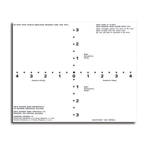
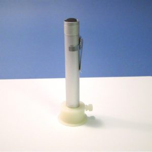 Phoria Cards Test vertical and horizontal muscle balance at near and far. Used with Maddox Rod and Penlight. Referral considerations indicated on cards for imbalance beyond certain prism diopters or balance beyond specified esophoria and exophoria ranges. Shows phoria to parents!Item #: BC1209+
 -
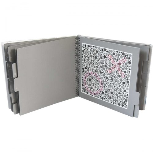 730006 The HRR (Hardy Rand and Rittler) Standard Pseudoisochromatic Test, 4th Edition provides several very important features to provide the most advanced color vision test available: congenital and acquired testing, identification of the type of defect, and diagnosis of the extent of the defect as well as quick positive classification of normals. The HRR employs a sophisticated test strategy that virtually eliminates the potential for memorization and malingering. The figures used by the HRR Pseudoisochromatic Plates are independent of language and suitable for both adults and children.
 -
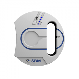
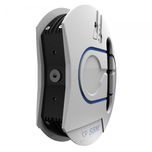 Typology: Device for evaluation of the meibomian glands Camera: Colored, sensitive to Infrared (NIR) Part examined: Upper and lower eyelids Graphic result: Coloration absent area and present Tools: Editor to highlight the area if the glands to be evaluated Resolution: 8 mp Light Source: Infrared LED Rating: Calculation of percentage of missing glands
 -
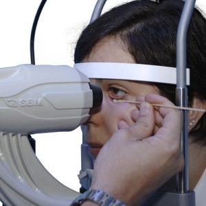
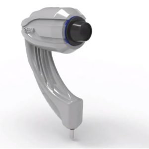 Acquisition Mode: Multi shot, tube, movie ISO Management: Variable Grids: Placido disc, NIBUT grid Light source: Infrared LED - Blue and White LED Image Resolution: 8 mp Focus: Autofocus, Manual focus Camera: Colored, Sensitive to Infrared (NIR)
 -
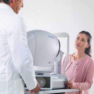
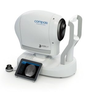
iCare COMPASS Automated Perimeter with active Retinal Tracking
Key features
- Standard automated perimetry
- Active retinal tracking compensating for poor patient fixation in real-time
- Auto-focus — no trial lens needed
- Hygienic design
- Illustrative fixation analysis; fixation area and plot
- High-resolution confocal TrueColor imaging of the retina
- No dilation of pupil needed, the patient can blink freely and the test can be suspended at any time without data loss
- Ease of use & minimal operator training
-
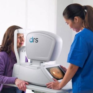
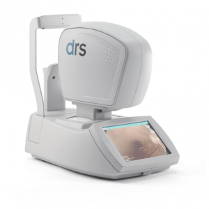
iCare DRS automated digital retinography system
Key features- Fully automated: patient autosensing, auto-alignment, auto-focus, auto flash adjustment, auto-capture
- Short exam time
- High quality color and Red-free images
- Minimal training required
- Compact and clean design
- No additional PC required
- Touch-screen operation
-
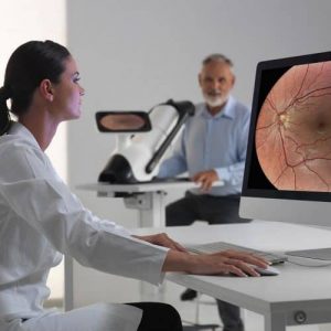
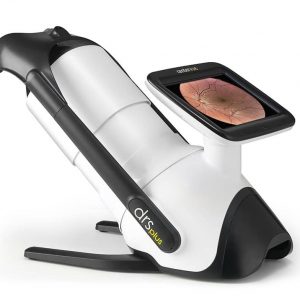
iCare DRSplus TrueColor confocal fundus imaging system
Key features- TrueColor Confocal Technology
- Multiple imaging modalities including red-free, external eye and stereo view imaging
- 2.5 mm minimum pupil size
- Fast, easy and fully automated operations
- Mosaic function which creates retinal panoramic views up to 80°
- Remote Viewer that allows for reviewing from devices on the same local area network
- Remote Exam feature enables executing an exam from a distance
-
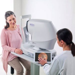
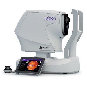
iCare EIDON widefield TrueColor Confocal fundus imaging system
Key features- Multiple imaging modalities including TrueColor, blue, red and Red-Free and infrared confocal images
- Widefield, ultra-high-resolution imaging
- Capability to image through cataract and media opacities
- Dilation-free operation (minimum pupil 2.5 mm)
- Flexibility of fully automated and fully manual mode
- All-in-one compact design, no additional PC required
-
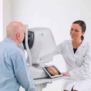
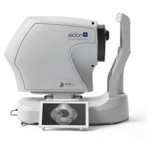
iCare EIDON AF blue autofluorescence confocal fundus imaging system
Key features
- TrueColor, blue autofluorescence, infrared and RGB channels confocal images
- Panoramic view of the retinal autofluorescence (up to 110°)
- High details and contrast in the same device
- Short exam time and enhanced patient comfort
- Easy to use, speeds up patient workflow
-
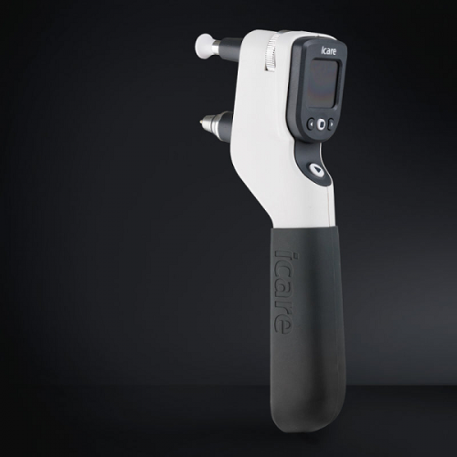
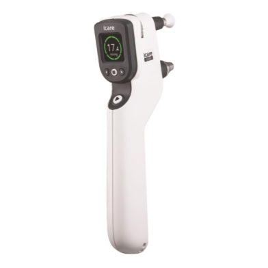
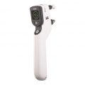
iCare IC200 tonometer - introducing a new era in clinical tonometry
Key features
- 200-degree position freedom
- Suitable for all patients
- Consistent and accurate readings
- No anesthetic drops
- Improved probe control
- User interface in multiple languages
- Wireless connection to iCare EXPORT
- Wireless printing
- No calibration
-
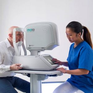
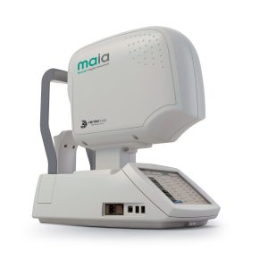
iCare MAIA and S-MAIA confocal microperimeters
Key features- 25 Hz eye-tracking with confocal SLO imaging
- Pre-programmed and custom tests
- Sensitivity and fixation indexes, illustrative sensitivity, and fixation maps
- Informative examination and progression reports
- 2.5 mm minimum pupil size
- Patients with cataract (up to grade 3+) and media opacities can be examined
- Auto-focus (from -15D to +10D)
- Sensitive to functional changes due to macular pathologies or treatments even in early stages (background down to <0.0001 asb, threshold range 36 dB)
- Simple to use, patients can be tested in less than 3 minutes per eye
- S-MAIA includes also scotopic testing with cyan and red stimuli
-
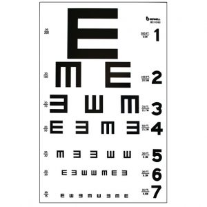 9" x 14" TRANSLUCENT PLASTIC MAY BE USED WITH ILLUMINATED TEST CABINET OR AS A WALL CHART 20/200 to 20/15. Item #: BC1262
 -
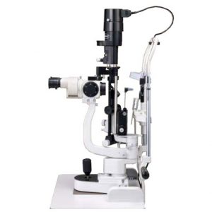 The L-0189 5 STEPS MAGNIFICATION SLIT LAMP provides practitioners with the utmost satisfaction thanks to its excellent image clearness and fine design.
 -
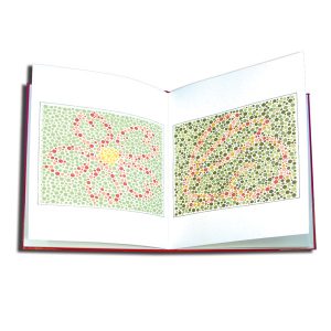 This test is similar to the Ishihara Isochromatic Test. 10 plates with non-verbal animal patterns. Originally created by Hiro Matsubara, MD. Printed in Japan. Item #: AT400

